The prostate > Anatomy of the prostate

Generalities and surrounding structures

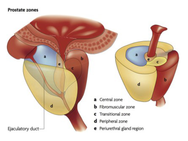
Located in the pelvis, the prostate stands below the bladder, behind the pubic bone, in front of the rectum and above the muscular pelvic floor. It is at the crossroads of the urinary and male reproductive systems. At 30 years, its normal size is 3 x 4 x 5 cm reaching a volume 25-30 mls often compared to a chestnut in size and shape (picture 2). It can also be compared to an inverted pyramid with the base above in contact with the bladder neck and the top below where the urethra comes out from the prostate. The upper part of the prostate is indeed called the base just below the bladder neck and the lower part the apex just above the urinary sphincter main structure acting for the urinary continence (picture 4). Eventually posteriorly on each side of the prostate run the neurovascular bundles essential to the activation and the maintenance of the erections (picture 5).

The urinary sphincter and the neurovascular bundles are so narrow to the prostatic gland that one can understand easily how any radical treatment such as the surgical removal or even extensive irradiation of the prostate will forcibly affect the continence and erectile functions.

picture 4

Picture 4 : Urinary sphincter immediately underneath the prostate (by B. Batard)

picture 5

Picture 5 : Neuro-vascular bundles (NVB) running posteriorly alongside the prostate. In 75% the NVBs are bilaterally well-delimited and dissectable, and in 25% the NVBs are spread and impossible to preserve. Before an ablation of the prostate, the NVBs anatomy is not predictable (by B. Batard)

The zonal description of the prostate

For decades the anatomical structure of the prostate has been perfectly described by Mc Neal. The urethra is always medial on the right/left axis whereas on the posterior/anterior axis it can be located at different levels. The picture 6 shows you a sagittal cross-sectional view of the prostate with the bladder neck above with a urethra quite central surrounded by the prostate. Beside the 3D prostate is constituted by different curved layers around a tube which is the urethra.

Immediately around the urethra 2 layers or zones are surrounding the urinary canal which are the transitional zone in blue and the central zone in brown (picture 7). The development of an adenoma of the prostate also called benign prostatic hyperplasia (BPH) is based on the increasing volume of the transitional zone with age starting for some men as early as 30 years. More generally for most of the male population and with some ethnic differences symptoms due BPH are more likely to appear after 50 years.

The second important zone of the prostate is located posteriorly recovering also laterally the gland in yellow in picture 7. It is called the peripheral zone. In 70-80% of the cases prostate cancer also called prostatic adenocarcinoma will take ground from this peripheral zone which explained the relevance of the digital rectal examination as almost sole diagnostic tool before the surge of the Magnetic Resonance Imaging.

The anterior zone in green completes the anatomical structure with the particularity of being deprived of prostatic glands (picture 8). It is also called the anterior fibro-muscular stroma.

Picture 6 : Zonal anatomy according to Mc Neal description

Picture 7 : Transversal cross sectional view of the different zones of the prostate

Picture 8 : 3D sagittal cross sectional view of the different zones of the prostate

The importance of the capsule

The prostatic gland is surrounded by a capsule which is an envelope delimitating entirely the gland except at certain points such as the posterior entry of the the ejaculatory ducts (video 1) or the exit of the urethra through the apex. This envelope has no special interest except for the prostate cancer staging with mainly the localized prostate cancer where the integrity of the capsule is totally respected and conversely a locally advanced cancer with a tumour going through the capsule and potentially invading adjacent structure such as the seminal vesicles, the rectum, the urinary sphincter or even the bladder. The disruption of the capsule by the tumour is not particularly related to the presence or absence of distant mestastasis.

A gland

Indeed the prostate is a gland constituted by glands, connective tissue and muscular fibers. The prostate tends to increase in size with age.

Role of male hormones

Male hormones are named androgens. Testosterone and Di-Hydrotestosterone (DHT) are amongst the best know amongst them.